Acquisition
- Use of PET camera is essential.
- 45-60 min after administration of 18F-FDG, acquire slices over 360 degrees for 10 min, 1 bed.
- Obtain a perfusion PET study for comparison with the metabolic images. 13N-NH4 and 15O-H2O are standard radiotracers, but not easily available. 18F Flurpiridaz is a possibility in the future, now under research.
- Alternatively it could be adequate to get a perfusion SPECT study with sestamibi for comparison with the metabolic PET images:
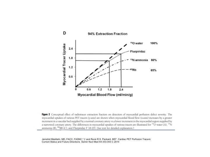
Thallium Spect or 99m Tc Sestamibi or 18F Flurpiridaz, note below its extraction fraction compared to other PET myocardial flow tracers.
References:
1 Gonzalez P et al. Viabilidad Miocardica, p 105-119 in: Medicina Nuclear Aplicaciones Clinicas. Eds: I. Carrio - P. Gonzalez. Editorial Masson, Barcelona España, 2003.
2 Bax JJ et al. 18-Fluorodeoxyglucose imaging with positron emission tomography and single photon emission computed tomography: cardiac applications. Semin Nucl Med. 2000 Oct;30(4):281-98.
3 Guehl NJ, et al. Single-scan rest/stress imaging: validation in a porcine model with (18)F-Flurpiridaz. Eur J Nucl Med Mol Imaging. 2017.