Miscellaneous Conditions. Prepared by Dr Gabriel Castro.
Duodenogastroesophageal reflux during
Technetium-99mSestamibi Cardiac Imaging
![]()
![]()
55 y.o female, with hypertension and smoking habit, with no prior coronary
disease backround. Underwent a Tc99m Sestamibi cardiac spect due to atypical
thoracic pain. She had history of prior surgery for achalasia, and
gastrosophagel reflux.
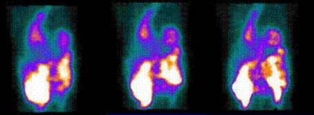
In raw
rotating images, Tc99m-sestamibi SPECT rest study shows activity throughout the
esophagus (yellow arrow), that also seems widened. In addition, marked activity
is seen in the stomach (white arrow). The patient referred an intense
gastroesophageal reflux episode during spect adquisition. Due to its
hepatobiliar excretion, we can appreciate radiotracer activity in stomach and
esophagus (Enterogastric Bile Reflux) (1). This is an
unusual finding, but a careful review of the raw rotating images should be an
integral part of the interpretation of all myocardial perfusion imaging studies
(2).
References:
1) Williams KA, Hill KA, Sheridan CM. Noncardiac findings on dual-isotope myocardial perfusion SPECT. J Nucl Cardiol 2003;10:395-402.
2)
Kabasak L et al. Enterogastric Bile Reflux during
Technetium-99mSestamibi Cardiac ImagingJ. Nucl Med 1996; 37:1285-128.
Home Index Myocardial Perfusion Spect Mibi Clinical Applications