Inflammatory Process (Prepared by Dr Guillermo Contreras)
79 year old male
patient with pain in the face and enlarged left parotid gland.
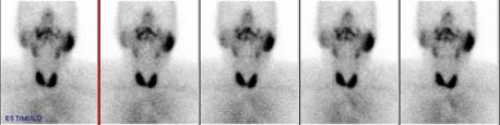
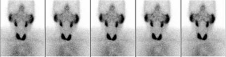
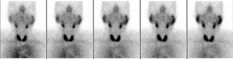
In the images, the left parotid gland appears enlarged and retains the
radiotracer.
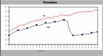
The excretion curves of the parotid glands, show the asymmetry between the
right (blue) which evidences a normal washout, and the left gland (red) which
reveals retention.
The patient recovered spontaneously and completely days later, suggesting an
inflamatory origin. However, this finding needs to be distinguished from a
salivary gland obstruction due to sialolithiasis.
References:
1 Roccia P, Di Liberto C, Speciale R, La Torretta G, Lo Muzio L, Campisi G. Obstructive sialoadenitis: update of diagnosis and therapy issues. Recenti Prog Med. 2006 May; 97(5): 272-9.
2 Klutmann S., Bohuslavizki K.H., Kroger S., Bleckmann C., Brenner W., Mester J. and Clausen M.. Quantitative Salivary Gland Scintigraphy. J Nucl Med Technol 1999; 27:20–26.