Gastroesophageal Reflux Scintigraphy Normal Images
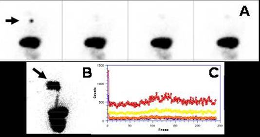
Boy 3 month old, presented with regurgitation. In A: Patient supine, images show a marker (arrow) at the cricoid cartilage. Stomach and bowel at the bottom. Sequential images during the 45 min acquisition did not reveal any activity increase in the esophagus. B: Arrow points to mouth when the patient was swallowing the liquid bolus, then the esophagus and stomach. C: Time activity curves over the esophagus. In red, total region of interest; yellow upper esophagus, orange middle and blue lower third.Curves are flat.
References:
1 Maurer AH, Parkman HP. Update on gastrointestinal scintigraphy.Semin Nucl Med. 2006 Apr;36(2):110-8.
2 Csendes A., González P., et al. Diagnóstico de Reflujo Gastroesofágico
mediante técnica isotópica. Rev Med Chile. 1989;117: 1374-1380.
Home Index Gastrointestinal Examinations Gastroesophageal Reflux Scintigraphy