![]()
Accessory Spleen
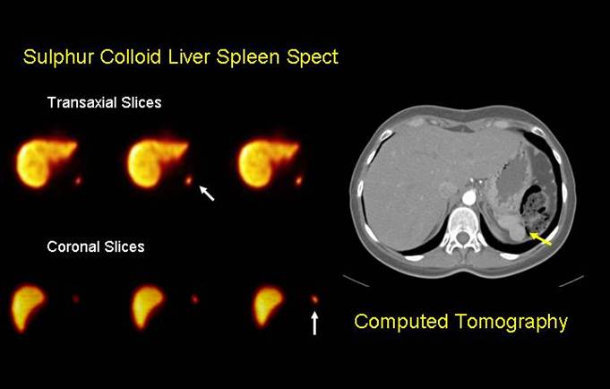
Twenty one year old female with history of Idiopathic Thrombocytopenic Purpura since 2001, splenectomy due to corticoid therapy failure. It Is admited with severe thrombocytopenia: 11000 platelets. Hepatoesplenic SPECT with 99mTc Sulphur Colloid shows accesory splenic tissue (white arrows), normal spleen not visualized. Note normal liver image. CT depicts 2 cm diameter accesory spleen adjacent to the pancreatic tail (yellow arrows). Prepared by Dr Gabriel Castro
Accessory spleen, a relatively common congenital defect, found in
10%-30% of patients at autopsy, is due to the fusion failure of the
splenic anlage, which is located in the dorsal mesogastrium. The splenic
hilus is the most common site of an accessory spleen followed by
pancreatic tail [
The most specific imaging method for diagnosing ectopic splenic tissue is nuclear scintigraphy [2]. Heat-damaged red blood cells have been shown to have a higher sensitivity and specificity than the sulfur colloid in the diagnosis of accessory spleen [3 ] but as demonstrated in this study, the ectopic splenic tissue was easily detected and localized with radiolabeled colloid
1 Freeman JL, Jafri SZ, Roberts JL, Mezwa DG, Shirkhoda A. CT of congenital and acquired abnormalities of the spleen. Radiographics. 1993;13:597–610.
2.2 Sica GT, Reed MF. Case 27: intrapancreatic accessory spleen. Radiology. 2000;217:134–137.
3.3 Accessory spleens in the thoracic and abdominal cavities after a relapse of idiopathic thrombocytopenic purpura: a case report. JK MacDonald, RA Wilke J Nucl Med Technol 2000; 28:49–51.