Intestinal Duplication (Collaboration of Dr Claudio Opazo)
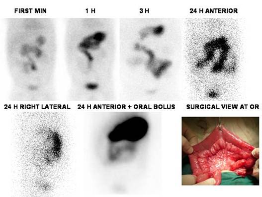
Male, 3 month old with lower GI bleeding. Underwent scan with 99mTc-Pertechnectate to rule out Meckel's Diverticulum. At the same time of stomach appearance, there is concentration in a quite atypical segment of the small bowel which persists up until 24 h. Six days later patient was operated on and a small bowel duplication was found and extracted. Clinical recovery was complete.
One segment of small intestine 23 cm long with a diameter ranging between 3 and 2.5 cm. From to 8.5 cm from one edge and 12.5 cm from the other edge there is an ovoid mass of 4 x 3.5 x 2.5 cm which corresponds to a cystic formation of 2x 1.5 x 1 cm. Peripheral fat congestive appearance is appreciated. Cecal appendix is 2.5 cm in length, without macroscopic changes, reaching a diameter of 4 mm, narrow lumen.
Histological signs are consistent with intestinal duplication cyst lining consisting of gastric mucosa composed of body type, with high cylindrical epithelium lining with high columnar cells with a fibrovascular stroma. The chorion contains lymphoid follicles and tubular normal glandular aspect. There is diffuse congestion and fibrinohematic intraluminal material.
Note high correlation between 24 h image (upper right frame) and surgical specimen shape at OR.
References:
Kiratli PO, Aksoy
T, Bozkurt MF, Orhan D. Detection of ectopic gastric mucosa using 99mTc
pertechnetate: review of the
literature. Ann Nucl Med. 2009 Feb;23(2):97-105.
Home Index Clinical Applications Meckel’s Diverticulum Diagnosis