Lymphoscintigraphy Normal Images (Prepared by Dr Isabel Berrocal)
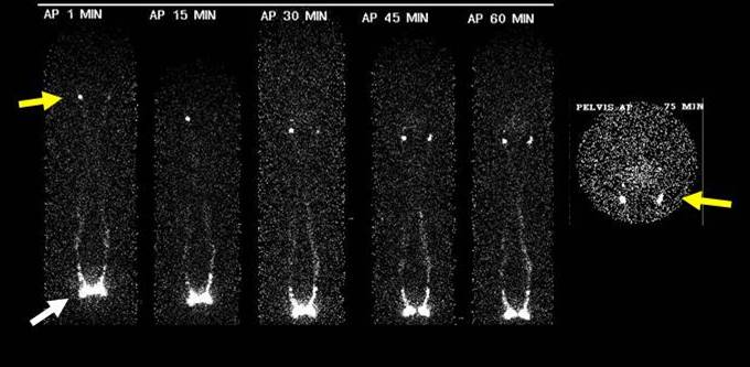
23 year- old female, with clinical suspicion of primary lymphedema of lower limbs. Isotopic lymphoscintigraphy was performed with 99m Tc NANOCINT, after intradermal injection with aseptic technique in the first interdigital spaces of feet (white arrow). Flow of the radiotracer is observed through the lymph vessels of both lower limbs, arriving in bilateral inguinal lymph nodes in early and late images, symmetrically (yellow arrow). Images were obtained at 1, 15, 30, 45, 60 and 75 min after injection.
Peripheral lymphatics can be easily visualized with radionuclide lymphoscintigraphy. It is an objective approach to diagnose and characterize the severity of lymphedema with a non invasive, repeatable procedure. Lymphangioscintigraphy can be performed before and after medical or surgical treatment and follow-up evaluation.
References:
1 Szuba A.,Shin W. The Third Circulation: Radionuclide Lymphoscintigraphy in the Evaluation of Lymphedema. J Nucl Med. 2003 Jan;44(1):43-57.
2 Williams W., Witte CL.Radionuclide lymphangioscintigraphy in the evaluation of peripheral lymphedema. Clin Nucl Med. 2000 Jun;25(6):451-64.