COPD
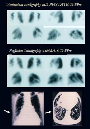
A 70 year old overinfected chronic pulmonary disease (mixted components) patient
in evaluation of pulmonary embolism is presented.
V/Q Scintigraphy shows reverse mismatched defects (bigger defects at ventilation
phase).
Chest x-ray and CT show opacity focci at right middle lobe and reticular
honeycomb infiltrate in right pulmonary base, compatible with fibrosis (arrows)
Prepared by: Dr
Alejandra Jiménez
Although more non diagnostic V/Q scan results can be expected in the presence of
COPD or other pulmonary diseases, it remains an informative, non invasive
screening.
V/Q Scan for diagnosis PE in COPD pacients has a Sensitivity of 79%,
Specificity of 92% (PPV:79%), (NPV:92%).
References:
1.- M.M.C. Tiel-van Buul, J.F. Verzijlbergen. Ventilation-Perfusion Lung
Scintigraphy.Imaging Decisions MRI 2004; 8 : 3-14.
2.- H.R. Ham et al. Ventilation-perfusion patterns in lung diseases ( with
reference to those observed in pulmonary embolism). Eur J Nucl Med 1985; 10:
165-166
Home Index 99 mTc-MAA Perfusion Scintigraphy 99 mTc Radioaerosol Ventilation Scintigraphy Clinical Applications