V/Q NORMAL IMAGES (Prepared by: Dr Alejandra Jiménez)
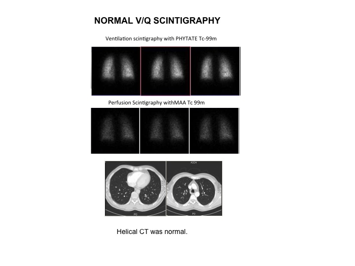
Male 46 year old with high risk for pulmonary embolism (PE).
Ventilation-Perfusion Scintigraphy Study shows homogeneous perfusion in the
parenchyma with mild irregularity at the hilum, and the same in ventilation
phase. There are no segmental or subsegmental defects. (Coronal slices).
Ventilation-Perfusion lung imaging is widely used as the initial screening
examination in the diagnosis of PE. This study has a high negative predictive value (NPV) : almost 100%. These is
no statistically significant overall diference in diagnostic perfomance between
helical CT and V/Q Scintigraphy
References:
1 McDonald WBG et al. Diagnosis of pulmonary embolism: Ventilation perfusion scintigraphy versus helical computed tomography pulmonary angiography. Australasian Radiology (2005) 49: 32-38.
2 Biello DR et al. Ventilation-Perfusion Studies in Suspected Pulmonary Embolism.AJR 1979; 133: 1033-1037.
Home Index Lung Imaging 99 mTc-MAA Perfusion Scintigraphy 99 mTc Ventilation Scintigraphy Normal Images