Pulmonary Aspiration Scintigraphy Normal Images
(Prepared by Dr Isabel Berrocal)
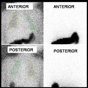
Male patient 1 year old, was referred for study of gastroesophageal reflux and lung aspiration. Dynamic study was performed with 99m-Tc sulfur colloid for 40 minutes and images for detection of pulmonary aspiration with and without transmission phantom and delay image, at 2 and 24 hours (the latter not shown). The study of reflux was positive with at least two marked episodes of tracer reaching the middle third of the esophagus of short duration. In chest imaging there is no evidence of abnormal activity in the bronchi or lungs.
Gastroesophageal scintigraphy and pulmonary aspiration images can be used to detect pulmonary aspiration during the postprandial period and it would be a strong evidence in favor of GER as a ethiologic factor. Usually during the test, the images are zoomed and centered on the lung fields and upper abdomen. A normal (ie, negative) overnight or late scan result is characterized by the absence of focal radioactivity within the lung fields.
References:
1 Ravelli M.; Panarotto M., et al. Pulmonary Aspiration Shown by Scintigraphy in Gastroesophageal Reflux-Related Respiratory Disease. CHEST 2006; 130:1520–1526.
2 Silver K., Van Nostrand D, et al. The Use of Scintigraphy in the Management of Patients with Pulmonary Aspiration. Dysphagia 9:107-115 (1994).