BRAIN VASCULITIS INFARCTS
Female patient 55 year old, with a history of cerebral vasculitis sequelae. EEG: delta theta slow continuous left temporo-occipital region without epileptiform activity. Bilateral internal carotid arteriography and left vertebral artery: regression of more than 95% arteritis signs of cerebral and cerebellar artery vessels compared with previous study. MR: Control of CNS vasculitis with bilateral brain infarcts with regressive changes. CT brain: Old lesions at occipital level, with greater involvement of right posterior frontal region and parasagittal frontal subcortical region precentral in the right side, in the context of CNS vasculitis.
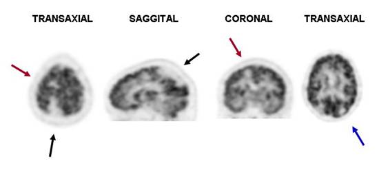
PET F18FDG Dose, 10 mCi, injected with glucose of 97 mg / dl.. Images show a large area of
decreased uptake of FDG in
right parieto-occipital lobes triangular shape and well defined edges (Black
Arrows). Additionally, there are two smaller areas, the first
high right parietal region at subcortical level (Red Arrows) and the second in the left
occipital area (Blue Arrow), both well-defined edges as well. The remaining brain scan, basal
ganglia and cerebellum with normaltracer distribution. Consistent with sequelae of vascular
events (infarcts).
Goedhart-de
Haan AM et al report a case with
vertebral artery vasculitis confirmed by 18-FDG
positron emission tomography, combined with CT angiography, and immediate
immunosuppressive therapy was started. Symptoms of stroke should, in a
particular clinical context, raise suspicion of giant cell arteritis.
References:
1 Quirce R. and Carril J.M. Spect y PET en la enfermedad cerebrovascular, p: 437-447; Mut F Demencias 453-464; Jimenez VA et al Tomografia por Emision de Positrones en el Diagnostico de los tumores del sistema nervioso central, p481-489 in: Medicina Nuclear Aplicaciones Clínicas. Eds: I. Carrio - P. González. Editorial Masson, Barcelona España, 2003.
2 Goedhart-de
Haan AM, Pans SJ, Lensen KD, Meijerink MR, Comans EF, Smulders YM. Vasculitis revealed by posterior stroke. Neth J Med. 2012
Mar;70(2):81-3.
Home Index Neuropsychiatric Procedures 18F-FDG Brain Imaging Clinical Applications