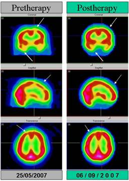
schyzophrenia
Male, 30 year old. Brain Spect with 99m Tc ECD, On 25/05/2007 shows heterogeneous perfusion in the brain cortex. Diminished frontal flow bilaterally and in left temporal lobe, with relative increased perfusion in the region of right temporal lobe (not shown in these slices). On 06/09/2007, after speciifc therapy, mild decreased perfusion in the left temporal lobe and appropriate activity in the rest of the brain cortex. (Arrows in saggital, transaxial and coronal slices).
Different abnormalities have been described in schizophrenia based on spect. Mainly decrease of perfusion in frontal and temporal lobes. During allucination phase, blood flow is augmented and the same occurs with radiotracer uptake in the corresponding cortex.
The first work done by these techniques put special emphasis on the 'hypofrontality' in schizophrenia. Likewise, this pattern of hypofrontality has been found during the course of certain cognitive tasks, such as the Wisconsin Card test, the Tower of London, etc. Regarding the temporal lobe, a review carried out in 2000, found increased temporal lobe activity in 13 SPECT studies and in 6 with PET. These techniques have also been employed to identify the areas involved in auditory hallucinations, and are associated with increased activity in the primary and secondary auditory cortex, regions of Broca and Wernicke, the striatum, the amygdala-hippocampal complex and the cingulate cortex.
References:
1 Cuevas-Esteban J, Campayo A, Gutiérrez-Galve L, Gracia-García P, López-Antón R. Background and findings of neuroimaging in schizophrenia: an update. Rev Neurol. 2011 Jan 1;52(1):27-36.
Home Index 99mTc-HMPAO Brain SPECT 99mTc-ECD Brain SPECT Clinical Applications