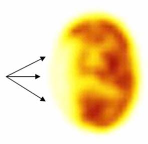
BRAIN INFARCT FDG
Boy, evaluated clinically at 2 months and at 6 years of age, bearing Takayasu's disease and arterial hypertension. When newborn, presented with sopor of abrupt onset. CT and MR (not shown) demonstrated a right extensive brain infarct. Patient recovered. Later on and to detect aortic active involvement, a FDG PET was requested. No abnormal uptake was noted in the arteries. Brain transaxial slice showed absence of metabolism in the right frontotemporoparietal cortex, compatible with infarct.
References:
1 Quirce R. and Carril J.M. Spect y PET en la enfermedad cerebrovascular, p: 437-447; Mut F Demencias 453-464; Jimenez VA et al Tomografia por Emision de Positrones en el Diagnostico de los tumores del sistema nervioso central, p481-489 in: Medicina Nuclear Aplicaciones Clínicas. Eds: I. Carrio - P. González. Editorial Masson, Barcelona España, 2003.
Home Index Neuropsychiatric Procedures 18F-FDG Brain Imaging Clinical Applications