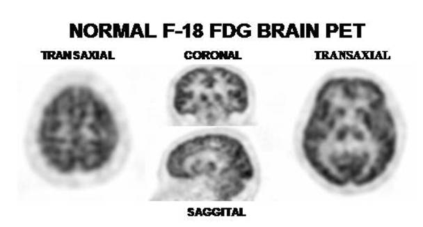
The images are from a 43 year old male complaining of memory deficit. Note homogeneous and symmetric FDG metabolism in both hemispheres. The slice to the left is passing near the vertex and the one to the right at the level of basal ganglia. It can be seen the caudate with the internal capsule and thalamus. In the coronal slice passing through the caudates, depicted are the brain cortex of the frontal and temporal lobes. The saggital slice is passing through the midline and displaying the central frontal, parietal and occipital gyrii. In this projection the cingulate cortex is nicely shown.
References:
1 Quirce R. and Carril J.M. Spect y PET en la enfermedad cerebrovascular, p: 437-447; Mut F Demencias 453-464; Jimenez VA et al Tomografia por Emision de Positrones en el Diagnostico de los tumores del sistema nervioso central, p481-489 in: Medicina Nuclear Aplicaciones Clínicas. Eds: I. Carrio - P. González. Editorial Masson, Barcelona España, 2003.
2 Van Heertum RL at al. 2-deoxy-fluorglucose-positron emission tomography imaging of the brain: current clinical applications with emphasis on the dementias. Semin Nucl Med. 2004 Oct;34(4):300-12.
Home Index Neuropsychiatric Procedures 18F-FDG Brain Imaging