Parkinson's Disease
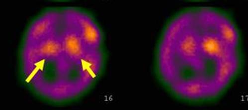
Female 65 year old with movement disorder more intense in the left arm. She underwent Trodat brain spect with 20 mCi (740 MBq) and 4 h after injection. Note bilateral uptake decrease more evident in the posterior nuclei and the right one.
Chou K.L. et al. using the posterior putamen as the main region of interest found the greatest accuracy (sensitivity 0.79, specificity 0.92). They concluded that TRODAT imaging can accurately differentiate early PD patients from controls and has the potential to improve the diagnosis of patients with early signs of PD (1).
Nonetheless, they recognize that [123I]b-CIT imaging was able to differentiate patients with PD from healthy controls with a sensitivity of 1.00 and a specificity of 0.95 (2).
Quantitative data can also be obtained and in several investigations demonstrate a significant difference between striatal uptake in control subjects and PD patients, especially obtained from the caudate and putamen. But in the individual case, a thorough visual assessment may be adequate enough to distinguish PD from other conditions.
References:
1 Chou K.L., Hurtig, HI., Stern, M.B., Colcher, A., Ravina,B., Newberg A., Mozley P.D., Siderowf, A.. Diagnostic accuracy of [99mTc]TRODAT-1 SPECT imaging in early Parkinson’s disease. Parkinsonism and Related Disorders 10 (2004) 375–379.
2 Parkinson Study Group, A multicenter assessment of dopamine transporter imaging with DOPASCAN/SPECT in parkinsonism. Neurology 2000;55(10):1540–7.