Bone Scan TemporoMandibular Disorder
(Prepared by Dr Isabel Berrocal)
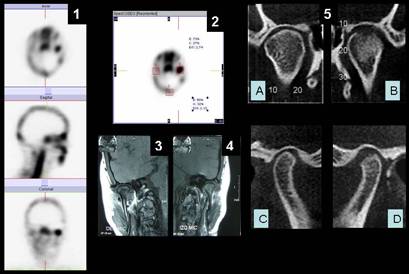
Female 24 y-o with
palpable TMJ bilateral pain and painful mouth-opening, underwent in 1: Tc-99m
MDP skull SPECT which showed intense osteoblastic activity in the left TMJ and
mild in the contralateral. In 2: TMJ indices vs themselves and vs skull were
consistent with inflammatory findings. 3 (right) and 4 (left): TMJ – MRI showed:
Anterior dislocation of both disks with chronic course in the left. Moderate
medial dislocation in the right joint and significantly left lateral dislocation.
Bilateral synovial inflammation more intense on left. 5: TMJ-CT showed: Figure
A, B front view : Both condyle cortex with an erosion area and osteosclerosis.
Right condyle (A) in central position and left condyle (B) in medial position
and beyond interarticularis space. Figure C, D side view: Both condyles in
posterior position. Left condyle (D) flattened. Image suggestive of arthritis.
References:
1.- Byeong-Cheol Ahn, et al. New quantitative method for bone tracer uptake of
temporomandibular joint using Tc-99m MDP skull SPECT. Ann Nucl Med (2009). Vol
23, Issue 7, pp: 651-656.13;
2.- Fahey Frederic H.Use of 99mTc-MDP SPECT for assessment of mandibular growth:
development of normal values. Eur J Nucl Med Mol Imaging (2010) 37:1002–1010
Home Index Clinical Applications Clinical Applications Inflammatory