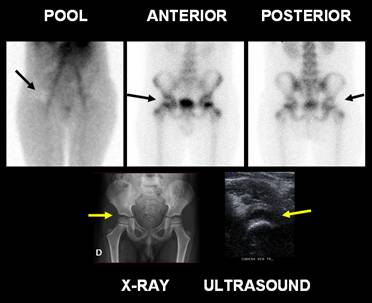
Bone Scan ACUTE SYNOVITIS
Five year old boy that presented at ER with pain in the right hip for a week duration.Ultrasound shows intra-articular effusion and synovial thickening. Blood pool phase shows avascular rounded area in the hip joint. In the delay projection there is increased joint space in correlation with the initial finding. X-ray, pelvis: Mild widening of the right hip joint space compared with left one. Sacroiliac joints and pubic symphysis without alterations. Bone scan demonstratesdecreased activity both at perfusion and delay phase, consistent with fluid in the joint, non specific and suggestive of an inflammatory process. The clinical course without fever was compatible with transient synovitis, blood cell count and PCR suggestive of a viral infection.
Gordon et al. evaluated 36 symptomatic children, using clinical findings, bone
scan, X-ray and follow-up, the children were divided into two groups: synovitis
or Perthes disease. In children with hip pain of over 1 week's duration, the
main value of the bone scan
is the early detection of Perthes disease. Diffuse increased activity on the
painful side suggests synovitis. A normal scan virtually excludes significant
skeletal abnormality.
References:
Gordon I, Peters
AM, Nunn R. The symptomatic hip in childhood: scintigraphic findings in the
presence of a
normal radiograph.Skeletal Radiol. 1987;16(5):383-6.
Home Index Clinical Applications Clinical Applications Inflammatory