Rheumatoid Arthritis
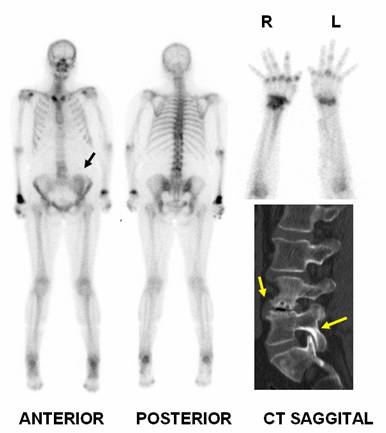
Female 54 year old, underwent kidney transplantation some years ago, currently normal renal function. Complains of joint pain in several sites mainly in pelvis. She was referred for a bone scan with diagnosis of reumathoid arthritis. Note severe wrists uptake and in various metacarpophalangeal joints (note ulnar fingers deviation), elbows, shoulders, right sternoclavicular junction, T10, L3-L4, knees, calcaneus at both sides. Sacroiliac Index was 1.3 in the two joints. Black arrow points to kidney transplant uptake in the left pelvis. No kidney visualization bilaterally in the renal lodge.
Lumbar spine CT performed after the bone scan was consistent with osteoarthritis mainly in spine (yellow arrows points to sclerotic lesions and gas in the vertebral body of L3).
Bone scan is sensitive but not specific to diagnose the cause of an inflammed joint. Thus, Gerasimou et al. using in 40 patients radionuclide imaging with human polyclonal immunoglobulin labeled with 99mTc, found an overall sensitivity of 83.3% and specificity at the level of 92%, being superior to MDP bone scan (1).
References:
1 Gerasimou G, Moralidis E, Papanastasiou E, Liaros G, Aggelopoulou T, Triantafyllidou E, Lytras N, Settas L, Gotzamani-Psarrakou A. Radionuclide imaging with human polyclonal immunoglobulin (Tc-HIG) and bone scan in patients with rheumatoid arthritis and serum-negative polyarthritis. Hippokratia. 2011 Jan;15(1):37-42.
Home Index Clinical Applications Clinical Applications Inflammatory