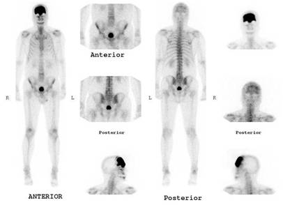
PAGET DISEASE SCAN
Male 59 year old,
Note diffuse increased 99m Tc MDP uptake, in the frontal bone, homogeneous. The
res of the skeleton depict mild osteoarthritis involvement in shoulders, lumbar
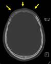
spine and knees. In the right hand side a CT slice fully correlated to the bone
scan with heterogenous increased density. No malignant sarcomatous findings seen.
(yellow arrows).
Paget disease of bone affects 3%-4% of the population over 40 years of age.
Three pathologic phases have been described: the lytic phase; the mixed phase;
and the blastic phase. Frequent sites of involvement include the skull
(25%-65%), spine (30%-75%) (mickey mouse sign), pelvis (30%-75%), and proximal
long bones (25%-30%). Bone scintigraphy demonstrates marked homogeneous increase
uptake in all phases of Paget disease. Complications include deformity and
fracture, arthritis, neurologic symptoms, and neoplastic involvement.
Sarcomatous transformation is the most feared complication, occurring in
approximately 1% of cases.
Smith SE, Murphey MD, Motamedi K, Mulligan ME, Resnik CS, Gannon FH.From the
archives of the AFIP. Radiologic spectrum of Paget disease of bone and its
complications with pathologic correlation. Radiographics. 2002
Sep-Oct;22(5):1191-216.