PLANAR Equilibrium
Radionuclide Ventriculography Normal Images
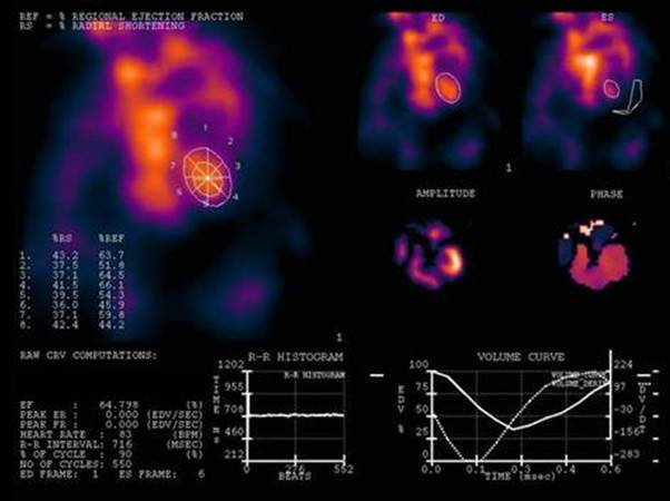
Female 68 y/o, referred to assess ventricular function previous to bone marrow transplantation due to multiple myeloma. Note in LAO normal size heart cavities. Nice separation in this view of right and left ventricle. Significant volume difference between end diastolic and end systolic frames (ED and ES, to the right hand side). Shown also amplitude and phase images pertaining to Fourier's processing. At bottom right, the volume curve. EF = 65% (normal over 52%). Heart rate was 83 bpm (R-R interval, 716 msec) and 550 cardiac cycles were acquired gating the R ECG wave. In vivo - in vitro RBC labeling with 99m-Tc was used.
A normal ejection fraction and regional wall motion contractility with adequate heart size at resting condition rules out ventricular function involvement.
Patient prognosis is excellent.
1 Russell RR 3rd, Zaret BL. Nuclear cardiology: present and future.Curr Probl Cardiol. 2006 Sep;31(9):557-629.
2 Kandora H, González P, Lillo R, Massardo T, Ortiz M, Asenjo R, Oyarzún R, Aramburu I, Loureiro O, Otárola S. Myocardial contraction and ventricular function in normal subjects evaluated with isotopic ventriculography and phase analysis. Rev Med Chil. 1991 Jul;119(7):733-8.
Home Index Cardiovascular Procedures Equilibrium Ventriculography