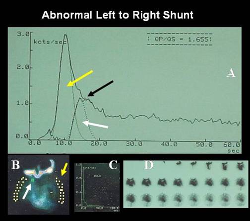
Male 18 year old patient, referred for left to right shunt evaluation. A ventricular septal defect was demonstrated on echocardiography. On A: Qp:Qs gave a value of 1.7 (Normal= 1.2). Yellow arrow, lung curve area; White arrow, shunt flow with early recirculation. Black arrow, recirculation peak. On B: regions of interest of superior vena cava for bolus quality control, in this case less than 2 sec (white arrow), both lungs (yellow arrow pointing to left lung roi). On D: Sequential images of first transit tracer bolus going through right and left cardiac cavities. Patient was injected on the left superficial yugular vein.
References:
1 Peix A. Medicina Nuclear en Cardiología Pediátrica, p 151-158 in: Medicina Nuclear Aplicaciones Clínicas. Eds: I. Carrio - P. González. Editorial Masson, Barcelona España, 2003.
2 Maltz DL, Treves S. Quantitative radionuclide angiocardiography: determination of Qp: Qs in children. Circulation. 1973 May;47(5):1049-56.